kfmrc@kau.edu.sa
00966-12-695-2000 Ext. (25027 ,61453)
Imaging Unit
The Imaging Unit (IU) located at the King Fahd Medical Research Center (KFMRC), King Abdulaziz University (KAU), Jeddah, Saudi Arabia (SA). The IU combines state-of-the-art instrumentation and a nationally recognized staff to assist investigators with a wide range of imaging based experimental approaches
This Facility can benefit any research group interested in using imaging techniques to further enhance scientific knowledge in any imaging related studies in the following areas: Oncology research, infectious disease, inflammation, metabolic diseases, neurology, gene therapy, stem cell biology, cardiovascular disease, immunology & transplantation biology, toxicology and drug metabolism studies. Furthermore, we offer support in the design of animal studies, the selection of suitable modalities, the processing of the raw data, as well as the evaluation/interpretation of the results and the publication of the study
Devices
Imaging and Radiology Equipments
Visual Sonics Vevo-2100 Ultrasound
The first high-frequency, high-resolution digital imaging platform with linear array technology and Colour Doppler Mode. Our Vevo2100 (FUJIFILM/VisualSonics) can produce very high visual and temporal resolution that makes it suitable for imaging small animals, from zebrafish to rats. The Vevo2100 system supports a wide range of preclinical research applications and is ideal for long-term studies
Research Applications
-
Cardiovascular research - precise visualisation and quantification of cardiac and vascular function, and blood flow analysis
-
Abdominal imaging - visualisation and assessment of function of various abdominal organs and systems including the kidneys, liver, urogenital and gastrointestinal systems
-
Cancer research - characterisation of tumours in vivo, monitoring of tumour development and responsiveness to therapy
-
Developmental research - visualisation and manipulation of embryos in utero
-
Ultrasound-guided injections - precise drug delivery to a particular organ, creation of orthotropic tumours and embryo injections
Bruker 9.4t Mri Animal Scanner
The Bruker 9.4T animal magnetic resonance imaging (MRI) system is a flexible and adaptable MRI scanner for preclinical imaging and research. The scanner is suitable for small animal studies, with the ability to image objects as large as 7cm in diameter and as small as 100 microns
Research Applications
-
Anatomical and functional MRI in neuroscience
-
Angiography in circulation
-
Relaxivity for molecular imaging
-
Cardiovascular (CV) profiling
-
Brain, Abdominal and Cardiovascular molecular imaging.
-
Muscular, Articular and Dynamic molecular imaging
-
Magnetic particle imaging (ex-vivo and in-vivo)
-
Soft tissue differentiation
-
Perfusion and metabolism functional MR imaging
-
Contrast enhanced imaging
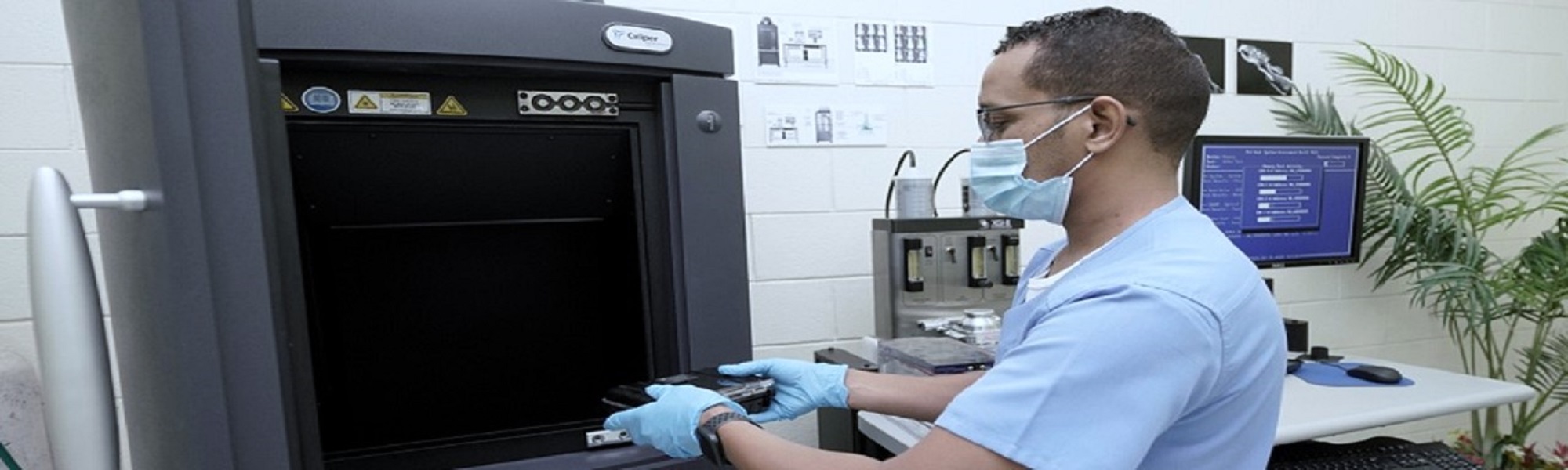
Caliper Ivis Spectrum In Vivo Imaging System
The IVIS Spectrum in vivo imaging system combines 2D optical and 3D optical tomography in one platform. The system uses leading optical technology for preclinical imaging research and development ideal for non-invasive longitudinal monitoring of disease progression, cell trafficking and gene expression patterns in living animals. It uses Xenogen’s novel patented optical imaging technology to facilitate non-invasive Bioluminescence or fluorescence longitudinal monitoring of disease progression, cell trafficking and gene expression patterns in living animals. The IVIS Spectrum pre-clinical optical imaging system combines high throughput and full tomographic capability for fluorescent and bioluminescent reporters
Research Applications
-
Advances in 3D Optical Imaging Quantification and Sensitivity
-
Cerenkov Imaging of Radioisotopes in IVIS systems
-
Stem Cell Research and Regenerative Medicine
-
Vascular Imaging Probes for Oncology and Inflammation Using the IVIS Spectrum
The Cellvizio 488 (Single Band)
Cellvizio LAB provides researchers with a new way to understand physiological phenomena at the cellular and sub-cellular levels in situ in animal subjects. Cellvizio Lab is easy to use and highly adapted for your research needs The equipment incorporates colour into the state of the art in vivo molecular imaging system. This allows researchers to visualize both invaluable anatomi- cal and functional information
Research Applications
-
Vasculature and vasodynamics studies
-
In vivo histology
-
Longitudinal monitoring of disease progression
-
Evaluation of cellular density
-
In vivo bio distribution imaging
-
Assessment of drug activity
Dr. Fahad Almutairi
Head of Imaging Unit
Mr. Mohammad Al-Nawar
Lab specilaist
Mr. Mohammed Assiri
Technichan
Mrs. Dalal Hanotoul
Technichan
Mr. Muferh Haressi
Technichan
Mr. Yahya Hull
Technichan
67334
Mr. Abdullah lasloom
Technichan
|
Last Update
1/8/2024 10:34:32 AM
|
|
|
|