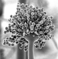
Effect of Aflatoxin B1 on colon morphology and motility on the mouse
|
|

Dr. Faiza Abdu, head of Neurology Unit at King Fahad Medical Research Center, pointed out that Aflatoxins B1 may represent a new model of post-inflammatory changes leading to gut dysfunction.
Her study revealed changes in colonic histology and ultrastructure following chronic exposure of AFB1 in mice and associated changes in colonic motor function. Aflatoxin b1 (20 and 40 ug/100g body weight) was administered orally in mice for 15 and 30 days of daily treatment. After 15 days, both doses caused shedding of mucosal epithelial cells together with hypertrophy and hyperplasia of goblet cells, inflammation of the lamina propria and an edematous muscularis propria. by 30 days, there was evidence of inflammation with infiltration of inflammatory cells and with the high dose there was an additional hypotrophy of the muscularis propria in both inner circular and outer longitudinal muscle. Ultrastructural abnormalities of nuclei and mitochondria were also observed.
In vitro segments of colon were mounted in a tissue bath and attached to pressure transducers to record intraluminal pressure changes indicative of peristaltic motor complexes (PMC). Neither dose had any effect on PMC amplitude or interval after 15 days treatment (p<0.05) in contrast, 30 days exposure resulted in a significant decrease in the amplitude (and interval) of PMC.
Dr. Abdu said that these findings indicate that chronic exposure to AFB1 induce inflammation and hypotrophy of the colon musculature accompanied be sever physiological changes in colon motility which contribute to gut dysfunction. Therefore, she recommends checking the foods infected with harmful fungus and to give blood samples for survey to ensure the absence of Aflatoxins B1.
|